

Being able to reproduce is a God-given gift. Those who have the ability to reproduce are said to be God's Creators. But how do we reproduce?
From Wikipedia, the free encyclopedia

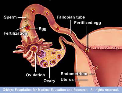
only one penetrates the ovum
Pregnancy starts with fertilization. Fertilization is the union of a humanoid egg and sperm, usually occurring in the ampulla of the uterine tube. The result of this union is the production of a zygote, or fertilized egg, initiating prenatal development. Scientists discovered the dynamics of human fertilization in the fourteenth century.
The process of fertilization involves a sperm fusing with an ovum—usually following ejaculation during copulation. It is possible, but less probable, for fertilization to occur without copulation, artificial insemination, or In vitro fertilization. Upon encountering the ovum, the acrosome of the sperm produces enzymes which allow it to burrow through the outer jelly coat of the egg. The sperm plasma then fuses with the egg's plasma membrane, the sperm head disconnects from its flagellum and the egg travels down the Fallopian tube to reach the uterus.
After fertilization, fetal development systematically and emidiately follows
Week 1–2
Gestational age: 0 to 1 (whole) weeks old. 1–14 days from last menstruation.
Embryonic age: -2 to -1 weeks old. (Week 1–2 of gestational age are theoretical extrapolations of embryonic age, since the fertilization hasn't actually occurred yet.)
Week 3
Gestational age: 2 (whole) weeks old. 15–21 days from last menstruation.
Embryonic age: Week nr 1. 0 (whole) weeks old. 1–7 days from fertilization.
- Fertilization of the ovum to form a new human organism, the human zygote. (day
- The zygote undergoes mitotic cellular divisions, but does not increase in size. This mitosis is also known as cleavage. A hollow cavity forms marking the blastocyst stage. (day 1.5–3 of fert.)
- The blastocyst contains only a thin rim of trophoblast cells and a clump of cells at one end known as the "embryonic pole" which include embryonic stem cells.
- The embryo hatches from its protein shell (zona pellucida) and performs implantation onto the endometrial lining of the mother's uterus. (day 5–6 of fert.)
- If separation into identical twins occurs, 1/3 of the time it will happen before day 5.
Week 4
Gestational age: 3 weeks old. 22–28 days from last menstruation.
Embryonic age: Week nr 2. 1 week old. 8–14 days from fertilization.
- Trophoblast cells surrounding the embryonic cells proliferate and invade deeper into the uterine lining. They will eventually form the placenta and embryonic membranes. The blastocyst is fully implanted day 7–12 of fert.
- Formation of the yolk sac.
- The embryonic cells flatten into a disk, two cells thick.
- If separation into identical twins occurs, 2/3 of the time it will happen between days 5 and 9. If it happens after day 9, there is a significant risk of the twins being conjoined.
- Primitive streak develops. (day 13 of fert.)
- Primary stem villi appear. (day 13 of fert.)
Week 5
Gestational age: 4 weeks old. 29–35 days from last menstruation.
Embryonic age: Week nr 3. 2 weeks old. 15–21 days from fertilization.
- A notochord forms in the center of the embryonic disk. (day 16 of fert.)
- Gastrulation commences. (day 16of fert.)
- A neural groove (future spinal cord) forms over the notochord with a brain bulge at one end. Neuromeres appear. (day 18 of fert.)
- Somites, the divisions of the future vertebra, form. (day 20 of fert.)
- Primitive heart tube is forming. Vasculature begins to develop in embryonic disc. (day 20 of fert.)


Week 6
Gestational age: 5 weeks old. 36–42 days from last menstruation.
Embryonic age: Week nr 4. 3 weeks old. 22–28 days from fertilization.
- The embryo measures 4 mm (1/8 inch) in length and begins to curve into a C shape.
- The heart bulges, further develops, and begins to beat in a regular rhythm. Septum primum appear.
- Branchial arches, grooves which will form structures of the face and neck, form.
- The neural tube closes.
- The ears begin to form as otic pits.
- Arm buds and a tail are visible.
- Pulmonary primordium, the first traits of the lung appear.
- Hepatic plate, the first traits of the liver appear.
- Buccopharyngeal membrane ruptures. This is the future mouth.
- Cystic diverticulum, which will become the gallbladder, and dorsal pancreatic bud, which will become the pancreas appear.
- Urorectal septum begins to form. Thus, the rectal and urinary passageways become separated.
- Anterior and posterior horns differentiate in the spinal cord
- Spleen appears.
- Ureteric buds appear.

Week 7
Gestational age: 6 weeks old. 43–49 days from last menstruation.
Embryonic age: Week nr 5. 4 weeks old. 29–35 days from fertilization.
- The embryo measures 9 mm (1/4 inch) in length.
- Lens pits and optic cups form the start of the developing eye.
- Nasal pits form.
- The brain divides into 5 vesicles, including the early telencephalon.
- Leg buds form and hands form as flat paddles on the arms.
- Rudimentary blood moves through primitive vessels connecting to the yolk sac and chorionic membranes.
- The metanephros, precursor of the definitive kidney, starts to develop.
- The initial stomach differentiation begins.
Week 8Gestational age: 7 weeks old. 50–56 days from last menstruation.
Embryonic age: Week nr 6. 5 weeks old. 36–42 days from fertilization.
- The embryo measures 13 mm (1/2 inch) in length.
- Lungs begin to form.
- The brain continues to develop.
- Arms and legs have lengthened with foot and hand areas distinguishable.
- The hands and feet have digits, but may still be webbed.
- The gonadal ridge begins to be perceptible.
- The lymphatic system begins to develop.
- Main development of external genitalia starts.
Week 9

Gestational age: 8 weeks old. 57–63 days from last menstruation.
Embryonic age: Week nr 7. 6 weeks old. 43–49 days from fertilization.
- The embryo measures 18 mm (3/4 inch) in length.
- Fetal heart tone (the sound of the heart beat) can be heard using doppler.
- Nipples and hair follicles begin to form.
- Location of the elbows and toes are visible.
- Spontaneous limb movements may be detected by ultrasound.
- All essential organs have at least begun
- The vitelline duct normally closes
Fetal period
The fetal period begins at the end of the 10th week of gestation (8th week of development). Since the precursors of all the major organs are created by this time, the fetal period is described both by organ and by a list of changes by weeks of gestational age.
Because the precursors of the organs are formed, fetus also is not as sensitive to damage from environmental exposures as the embryo. Instead, toxic exposures often cause physiological abnormalities or minor congenital malformation.
Changes by organ
Each organ has its own development.
- Development of circulatory system
- Heart development
- Development of digestive system
- Development of endocrine system
- Development of integumentary system
- Development of lymphatic system
- Development of muscular system
- Development of nervous system
- Development of the urinary and reproductive system
- Development of respiratory system
Changes by weeks of gestation
From the 8th week until birth (around 38 weeks), the developing organism is called a fetus. The fetus is not as sensitive to damage from environmental exposures as the embryo, and toxic exposures often cause physiological abnormalities or minor congenital malformation. All major structures are already formed in the fetus, but they continue to grow and develop.

Weeks 10–12
Gestational age: 9–11 weeks old.
Embryonic age: Weeks nr 8–10. 7–9 weeks old.
- Embryo measures 30–80 mm (1.2–3.2 inches) in length.
- Ventral and dorsal pancreatic buds fuse during the 8th week
- Intestines rotate.
- Facial features continue to develop.
- The eyelids are more developed.
- The external features of the ear begin to take their final shape.
- The head comprises nearly half of the fetus' size.
- The face is well formed
- The eyelids close and will not reopen until about the 28th week.
- Tooth buds, which will form the baby teeth, appear.
- The limbs are long and thin.
- The fetus can make a fist with its fingers.
- Genitals appear well differentiated.
- Red blood cells are produced in the liver.
Weeks 13 to 16
Gestational age: 12–15 weeks old.
Embryonic age: Weeks nr 11–14. 10–13 weeks old.
- The fetus reaches a length of about 15 cm (6 inches).
- A fine hair called lanugo develops on the head.
- Fetal skin is almost transparent.
- More muscle tissue and bones have developed, and the bones become harder.
- The fetus makes active movements.
- Sucking motions are made with the mouth.
- Meconium is made in the intestinal tract.
- The liver and pancreas produce fluid secretions.
- From week 13, sex prediction by obstetric ultrasonography is almost 100% accurate.
- At week 15, main development of external genitalia is finished

Week 20
Gestational age: 18 weeks old.
Embryonic age: Week nr 17. 16 weeks old.
- The fetus reaches a length of 20 cm (8 inches).
- Lanugo covers the entire body.
- Eyebrows and eyelashes appear.
- Nails appear on fingers and toes.
- The fetus is more active with increased muscle development.
- "Quickening" usually occurs (the mother and others can feel the fetus moving).
- The fetal heartbeat can be heard with a stethoscope.
Week 23
Gestational age: 22 weeks old.
Embryonic age: Week nr 21. 20 weeks old.
- The fetus reaches a length of 28 cm (11.2 inches).
- The fetus weighs about 925g.
- Eyebrows and eyelashes are well formed.
- All of the eye components are developed.
- The fetus has a hand and startle reflex.
- Footprints and fingerprints continue forming.
- Alveoli (air sacs) are forming in lungs.
Week 27
Gestational age: 26 weeks old.
Embryonic age: Week nr 25. 24 weeks old.
- The fetus reaches a length of 38 cm (15 inches).
- The fetus weighs about 1.2 kg (2 lb 11 oz).
- The brain develops rapidly.
- The nervous system develops enough to control some body functions.
- The eyelids open and close.
- The cochleae are now developed, though the myelin sheaths in neural portion of the auditory system will continue to develop until 18 months after birth.
- The respiratory system, while immature, has developed to the point where gas exchange is possible.
Week 31
Gestational age: 30 weeks old.
Embryonic age: Week nr 29. 28 weeks old.
- The fetus reaches a length of about 38–43 cm (15–17 inches).
- The fetus weighs about 1.5 kg (3 lb 0 oz).
- The amount of body fat rapidly increases.
- Rhythmic breathing movements occur, but lungs are not fully mature.
- Thalamic brain connections, which mediate sensory input, form.
- Bones are fully developed, but are still soft and pliable.
- The fetus begins storing a lot of iron, calcium and phosphorus
Week 35
Gestational age: 34 weeks old.
Embryonic age: Week nr 33. 32 weeks old.
- The fetus reaches a length of about 40–48 cm (16–19 inches).
- The fetus weighs about 2.5 to 3 kg (5 lb 12 oz to 6 lb 12 oz).
- Lanugo begins to disappear.
- Body fat increases.
- Fingernails reach the end of the fingertips.
- A baby born at 36 weeks has a high chance of survival, but may require medical interventions.

Weeks 36 to 39
Gestational age: 35–38 weeks old.
Embryonic age: Weeks nr 34–37. 33–36 weeks old.
- The fetus is considered full-term at the end of the 37th week of gestational age.
- It may be 48 to 53 cm (19 to 21 inches) in length.
- The lanugo is gone except on the upper arms and shoulders.
- Fingernails extend beyond fingertips.
- Small breast buds are present on both sexes.
- Head hair is now coarse and thickest.
The development is continued postnatally with adaptation to extrauterine life and child development stages.

See how magnificent the process is?
This is an example of God's love for us. By sharing His power to reproduce, we are able to experience how a creator loved his creation. Every parent would says that they will do every thing for their child. So as God.. God will always want the best for us and He will provide everything that is best for us. Although others may not feel it because they are having great problem, still in every situation God's Holiness is always present. We just have to remember that He have His reasons why we experience some difficulties.
As a parent, we should nurture and treasure the gift that God had intrusted to us.Take good care of them as much as we can and let us expose them in all aspect that can provide them growth specially Spiritually. Let us be thier guide to the journey that they will choose to take.
References:
http://images.search.yahoo.com/images/view;_ylt=A2KJke10WKVO11UA1XmJzbkF;_ylu=X3oDMTBlMTQ4cGxyBHNlYwNzcgRzbGsDaW1n?back=http%3A%2F%2Fimages.search.yahoo.com%2Fsearch%2Fimages%3Fp%3Dfetal%2Bdevelopment%26fr2%3Dpiv-web%26b%3D29%26tab%3Dorganic&w=588&h=248&imgurl=pregnancyearlysigns.org%2Fwp-content%2Fuploads%2F2011%2F07%2FFetal-Development.jpg&rurl=http%3A%2F%2Fpregnancyearlysigns.org%2Ffetal-development-month-to-month.html&size=35.3+KB&name=Fetal+Development+300x121+Fetal+Development+Month+To+Month&p=fetal+development&oid=05aa1955d8fb0898dd6339cd07e0daa4&fr2=piv-web&fr=&tt=Fetal+Development+300x121+Fetal+Development+Month+To+Month&b=29&ni=28&no=35&tab=organic&sigr=124udnslk&sigb=12t961rkq&sigi=128aeps61&.crumb=EJYbcsMi2eD
http://en.wikipedia.org/wiki/Fetal_development
http://en.wikipedia.org/wiki/Fertilization